CPPD Pseudogout: Why Calcium Crystals Mimic Arthritis
A swollen knee in an older adult can look like gout, rheumatoid arthritis, septic arthritis, or a mechanical flare. When the culprit is calcium pyrophosphate deposition disease (CPPD), the diagnostic challenge is that crystals create inflammation that closely resembles other forms of arthritis. That resemblance matters: CPPD is common, underrecognized, and often managed differently from autoimmune disease despite sharing the same clinical stage: a hot, painful, function-limiting joint.
For patients, the question is often, “Why did I get arthritis if I don’t have rheumatoid arthritis?” For clinicians, the question is practical: how do we confirm CPPD, exclude infection, and treat inflammation without committing someone to unnecessary long-term immunosuppression?
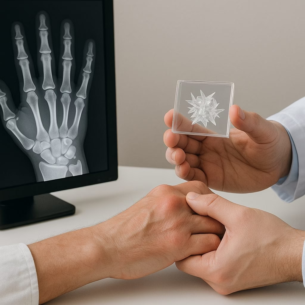
What CPPD Is, and Why It Behaves Like Inflammatory Arthritis
CPPD is caused by calcium pyrophosphate crystals forming in cartilage and related joint tissues. Unlike monosodium urate crystals in gout, CPP crystals are not primarily a blood-level problem that can be followed with a serum target. They reflect local cartilage biology, aging, prior joint injury, and sometimes metabolic contributors such as hemochromatosis, hyperparathyroidism, hypomagnesemia, or hypothyroidism.
The immune system reacts to deposited crystals as danger signals. Macrophages activate the NLRP3 inflammasome, a molecular alarm that increases interleukin-1 beta, a potent inflammatory cytokine. Neutrophils then flood the joint, producing warmth, swelling, and severe pain. This is why CPPD can look indistinguishable from gout at the bedside and can even resemble a rheumatoid arthritis flare when wrists, metacarpophalangeal joints, or knees are involved.
Terminology matters
“Pseudogout” describes the acute gout-like attack, but CPPD disease is broader. EULAR terminology separates acute CPP crystal arthritis, chronic CPP inflammatory arthritis, osteoarthritis with CPPD, and asymptomatic chondrocalcinosis seen on imaging. That distinction prevents a common misconception: calcium deposits on an x-ray do not automatically explain every ache. The clinical pattern and inflammatory findings still matter.

How CPPD Is Diagnosed Without Overcalling Arthritis
The most decisive test is synovial fluid analysis from the affected joint. Under compensated polarized light microscopy, CPP crystals are typically rhomboid and weakly positively birefringent. That wording sounds technical, but the principle is simple: seeing the crystal in inflammatory joint fluid connects the patient’s symptoms to a specific mechanism. Fluid analysis also allows cell count, Gram stain, and culture when infection is possible.
The 2023 ACR/EULAR classification criteria for CPPD disease strengthened research consistency by combining clinical features, metabolic associations, imaging findings, and crystal confirmation. Classification criteria, however, are not identical to bedside diagnosis. In practice, rheumatologists weigh probability: age, joint distribution, attack tempo, radiographic chondrocalcinosis, ultrasound findings, and whether another explanation better fits.
A clinical scenario
Consider a 72-year-old man with abrupt knee swelling two days after hospitalization for pneumonia. The knee is warm, tense, and painful with range of motion. His serum urate is normal. Aspiration shows inflammatory fluid, no organisms on Gram stain, and CPP crystals. The diagnosis is not “arthritis in general”; it is crystal-driven inflammation triggered in a vulnerable joint during systemic stress.
Why CPPD Gets Confused With Rheumatoid Arthritis and Gout
CPPD can mimic rheumatoid arthritis because it may become chronic and polyarticular, especially in the wrists and hands. It can cause morning stiffness, elevated C-reactive protein, and recurrent synovitis. The distinction is important because rheumatoid arthritis is an adaptive immune disease often associated with anti-CCP antibodies and erosive progression, while CPPD is innate immune activation around crystals. Disease-modifying antirheumatic drugs used for rheumatoid arthritis generally do not dissolve CPP crystals.
CPPD also mimics gout, but the chemistry differs. Gout management includes sustained urate-lowering therapy when indicated because urate is measurable and modifiable. CPPD has no equivalent proven crystal-depleting therapy. A normal uric acid level during an acute attack does not exclude gout, and chondrocalcinosis does not prove CPPD is the active problem; aspiration resolves that ambiguity when feasible.
Treatment Reasoning: Suppress Inflammation, Respect Comorbidity
Acute CPPD treatment targets inflammation rather than crystal removal. EULAR management recommendations support joint aspiration and intra-articular glucocorticoid injection for accessible monoarthritis, with oral NSAIDs, colchicine, or systemic corticosteroids considered based on kidney function, anticoagulation, diabetes, infection risk, and frailty. This is clinical reasoning, not a menu. The best choice is often the one that calms synovitis while creating the least collateral risk.
Common treatment considerations
- Joint aspiration can reduce pressure and provides diagnostic fluid.
- Intra-articular steroid may limit systemic exposure when infection is excluded.
- Low-dose colchicine may reduce recurrent attacks, although gastrointestinal, renal, and drug-interaction risks matter.
- IL-1 blockade has biologic plausibility and case-series support for selected refractory cases, but evidence is less mature than in gout.
Chronic CPPD inflammatory arthritis is harder. Small studies and expert practice support cautious use of colchicine, low-dose corticosteroids, or selected conventional agents such as methotrexate or hydroxychloroquine in persistent inflammatory phenotypes, but results are mixed. The uncertainty is the point: treatment should match demonstrable inflammation, not radiographic calcification alone.
What the Evidence Supports Now
The evidence supports three firm conclusions. First, CPPD is a true inflammatory arthritis caused by calcium pyrophosphate crystals, not merely “wear and tear.” Second, crystal identification in synovial fluid remains the most persuasive diagnostic anchor when the presentation is acute, atypical, or high stakes. Third, treatment is anti-inflammatory and individualized because no therapy has yet been proven to reliably remove CPP crystals from joints.
Uncertainty remains about prophylactic colchicine, chronic CPPD phenotypes, and selective cytokine inhibition. CPPD deserves precision because its mimicry is not harmless; accurate diagnosis prevents undertreatment of inflammation and unnecessary overtreatment as autoimmune disease.
Medically reviewed by Dr. Adam Elisha, DO, board-certified rheumatologist in Duluth, MN.