Why pseudogout deserves a careful look
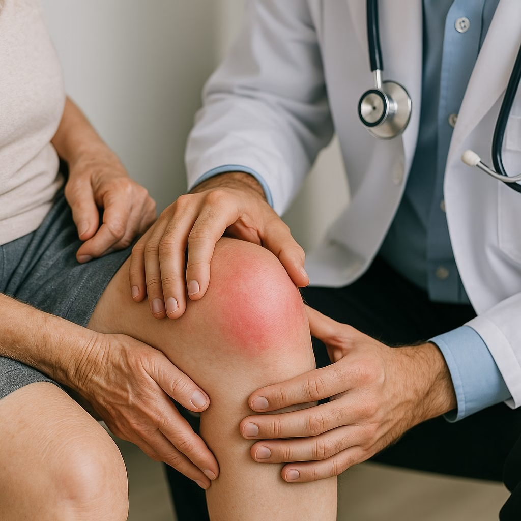
An older adult with a hot, swollen knee is often labeled as having gout, but that shortcut can mislead care. Pseudogout, now usually discussed under the broader term calcium pyrophosphate deposition disease (CPPD), can look nearly identical at the bedside. The distinction matters because the crystals, triggers, associated metabolic conditions, and long-term management differ. For patients, the question is why this joint suddenly became inflamed. For clinicians, the question is whether the episode represents infection, monosodium urate gout, CPPD, or another inflammatory arthritis.
Evidence supports a pragmatic approach: identify the crystal when possible, treat the inflammatory flare promptly, and look for patterns that change prevention. The 2011 EULAR recommendations for CPPD management remain a useful framework, emphasizing joint aspiration for diagnosis and anti-inflammatory therapy tailored to age, kidney function, bleeding risk, and comorbidities.

What happens in CPPD
Pseudogout occurs when calcium pyrophosphate crystals form in cartilage or nearby tissues and spill into the joint space. The immune system recognizes these crystals as danger signals, activates inflammatory pathways including the NLRP3 inflammasome and interleukin-1, and produces the familiar flare: heat, swelling, pain, and limited motion. Unlike rheumatoid arthritis, CPPD is not primarily an autoimmune disease; it is a crystal-associated inflammatory arthritis that becomes more common with aging.
Many patients have chondrocalcinosis, meaning calcification visible within cartilage on radiographs, especially in knees, wrists, and hips. Chondrocalcinosis supports the diagnosis, but it is not definitive: some people with deposits never flare, and some with true CPPD have subtle imaging. Polarized light microscopy remains the diagnostic anchor, showing weakly positively birefringent, rhomboid-shaped calcium pyrophosphate crystals in synovial fluid.
Symptoms patients and clinicians should recognize
An acute CPPD flare typically develops over hours to a day. The knee is the classic site, but wrists, ankles, elbows, shoulders, and the first metatarsophalangeal joint can be involved. Fever and elevated C-reactive protein can occur, which is why septic arthritis must remain on the table until the clinical picture and, when needed, synovial fluid studies argue otherwise.
A common scenario is a 74-year-old who wakes with a large, painful knee effusion two days after hospitalization for pneumonia. The knee is warm and difficult to bend. Laboratory testing shows leukocytosis, and the X-ray notes chondrocalcinosis. Aspiration finds inflammatory fluid with CPP crystals and negative Gram stain. That sequence illustrates typical reasoning: do not assume gout; prove the crystal pattern and protect against missing infection.
Causes and risk factors
Age is the strongest risk factor. CPPD is uncommon before age 60, then rises steadily as cartilage biology changes. Osteoarthritis and prior joint injury also create local environments where crystals are more likely to form. Flares may follow acute illness, surgery, trauma, or rapid shifts in fluid and electrolytes, which helps explain hospital-associated presentations.
When CPPD appears unusually early, is recurrent without obvious explanation, or has extensive joint involvement, clinicians often screen for associated metabolic conditions. These include hemochromatosis, hyperparathyroidism, hypomagnesemia, hypothyroidism, and chronic kidney disease. The reason is not academic: finding iron overload or low magnesium, for example, may change broader medical management, even though removing the association does not reliably erase existing crystals.
How pseudogout differs from gout
The names invite confusion. “Pseudogout” means “false gout,” but CPPD is not a minor version of gout. Both cause abrupt inflammatory arthritis, and both can coexist in the same patient. The difference is the crystal chemistry and the clinical context.
Gout management has a clear treat-to-target strategy in appropriate patients: lower serum urate below saturation, commonly below 6 mg/dL, using urate-lowering therapy when guideline indications are met. The 2020 American College of Rheumatology gout guideline strongly supports this approach for patients with tophi, radiographic damage, or frequent flares. CPPD has no comparable medication that dissolves calcium pyrophosphate crystals. Treatment therefore focuses on flare control, preventing predictable recurrences, and addressing contributors.
Diagnosis: why aspiration still matters
Serum uric acid is often overused as a shortcut. A high value does not prove gout, and a normal value during a flare does not exclude it. Likewise, chondrocalcinosis does not prove the painful joint is inflamed because of CPPD. Synovial fluid analysis answers several questions at once: white cell count, culture when infection is a concern, and crystal identification.
Ultrasound and radiography can strengthen suspicion, especially when aspiration is not feasible. The 2023 ACR/EULAR CPPD classification criteria formalized combinations of crystal proof, imaging findings, clinical pattern, and associated conditions for research classification. Classification criteria, however, are not the same as bedside diagnosis; clinical judgment remains essential.
Treatment: controlling inflammation without overreaching
Acute treatment is anti-inflammatory. Options include joint aspiration with intra-articular glucocorticoid injection for one or two accessible joints, short courses of oral glucocorticoids, low-dose colchicine, or nonsteroidal anti-inflammatory drugs in carefully selected patients. The choice depends less on elegance and more on safety: kidney disease, anticoagulation, heart failure, diabetes, ulcer history, and infection risk all change the calculation.
For recurrent flares, EULAR supports considering prophylactic low-dose colchicine when tolerated. In severe, refractory CPPD, small studies and case series have explored interleukin-1 blockade, but evidence is limited and patient selection matters. Chronic CPPD inflammatory arthritis can resemble seronegative rheumatoid arthritis, so treatment decisions should be anchored in distribution, imaging, synovial findings, and response over time.
Misconceptions and the current bottom line
The key misconception is that pseudogout is simply gout with normal uric acid. Current evidence supports crystal confirmation, individualized anti-inflammatory treatment, and uncertainty about disease-modifying therapy.
Medically reviewed by Dr. Adam Elisha, DO, board-certified rheumatologist in Duluth, MN.